Research Highlights
Kakshine, an unprecedentedly versatile new DNA staining probe
The research group of Kakishi Uno, Nagisa Sugimoto and Yoshikatsu Sato, of the Nagoya University Institute of Transformative Bio-Molecules (WPI-ITbM), has developed Kakshine, a new fluorescent staining probe for DNA suitable for use in the vast majority of organisms.
DNA contains the data that organisms pass on to their offspring. In eukaryotic organisms, DNA is mostly contained within the cell nucleus, with the mitochondria also possessing their own DNA. DNA fluorescent reagents exist for daily use in molecular biology such as electrophoresis and PCR, and are also indispensable for bioimaging analysis of chromosome dynamics and organelle replication during the cell cycle.
The ideal DNA fluorescent dye should have the following three qualities:
1) A high selectivity for DNA
2) Be applicable to visible light with low phototoxicity
3) Be applicable to a wide range of organisms
However, no dye that meets all of these requirements had yet been created. Kakshine, the new probe developed by this group, not only meets these three requirements, it adds the possibility for use in deep tissue imaging using two-photon excitation microscopy and super high-resolution STED live imaging of both nuclear DNA and organelle DNA, and is expected to find use at the cutting edge of life science research.
This research was published on May 11, 2021, in the journal Nature Communications.
About the research
DNA (deoxyribonucleic acid) is the carrier of an organism's genetic information, and DNA fluorescent reagents are an essential molecular tool in life science research. In cell biology, in particular, the dynamic analysis of DNA in living cells has been drawing attention using the very latest imaging techniques. However, conventional DNA dyes do not possess the qualities required for this level of research. Those which possess high selectivity for DNA require the use of phototoxic UV light, and those which can be applicable to the visible light spectrum possess insufficient DNA selectivity. To develop a new dye which would allow them to solve both of these problems at once, the research group set about creating a new molecule with different characteristics to existing DNA dyes. In the process, Kakishi Uno, who was studying for his PhD at the time, focused on the pyrido cyanine backbone, which had not previously been reported as a DNA dye. As a result of repeated improvements to this structure, he succeeded in synthesizing a derivative which allowed the dyeing of DNA with a fluorescence in the long-wavelength visible light spectrum (Figure 1). In honour of his discovery, and the fact that 'nucleus' in Japanese is pronounced 'kaku', the research group gave the series of new molecules, whose properties come from the symmetrical pyrido cyanine derivative, the name 'Kakshine'.
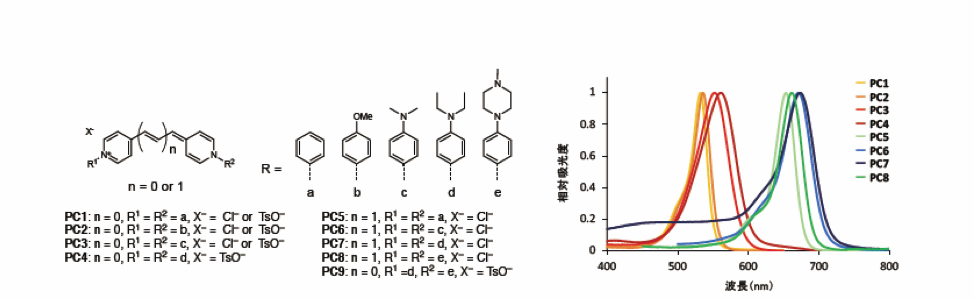
Figure 1: The structures of Kakshine series and their absorption spectra
By changing the length of the N-aryl group and methine chain, the research group were able to create a variety of compounds which would absorb the long-wavelength region of visible light (500-700 nm). All of these compounds were found to enhance their fluorescent intensities when bound to double-stranded DNA.
The group showed that the basic structure of Kakshine, PC1 (N-aryl Pyrido Cyanine Dye 1), specifically binds to DNA Adenine (A) and Thymine (T) double-stranded sequence regions, greatly increases the intensity of its fluorescence, and has a greater selectivity for DNA than the often-used commercial dyes, Hoechst and Pico-Green (Figure 2). As large amounts of RNA also exist within cells, it is particularly important that DNA dyes have a high selectivity for DNA, and the high selectivity of Kakshine highlights its usefulness.
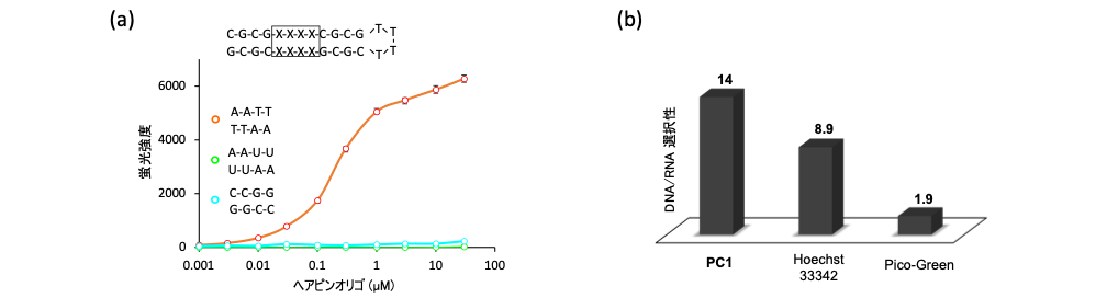
Figure 2: Binding properties of Kakshine to double-stranded DNA and its high DNA/RNA selectivity
(a) Kakshine (PC1) enhances its fluorescence in a concentration-dependent manner only for hairpin oligonucleotides containing AT sequences.
(b) The selectivity for DNA vs RNA of Kakshine compared to other reagents demonstrates its high performance.
Next, they showed that Kakshine could stain DNA in living plant cells as well as cultured animal cells (Figure 3). Kakshine's ability to penetrate right to the deepest parts of plant roots and be observable by two-photon excitation microscopy was particularly notable.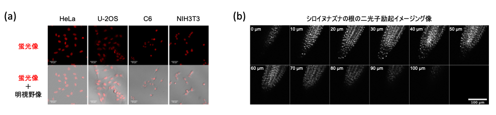
Figure 3: Live imaging of nuclear DNA with Kakshine
Kakshine was shown to be able to penetrate and stain nuclei in cultured animal cells including mouse, rat and human, and plant cells. Image (b) shows two-photon excitation imaging of root cells in Arabidopsis thaliana. Kakshine was able to penetrate right to the deepest regions of the root.
Next, the group showed that Kakshine can be used to stain either nuclear DNA or organelle DNA depending on its concentration. In human HeLa cultured cells, a concentration of 10 nM specifically stains nuclear DNA (nu-DNA), and a concentration of 100 pM specifically stains mitochondrial DNA (mt-DNA) (Figure 4a). Moreover, at the concentration at which both are stained, 1 nM, they succeeded in differentiating between nuclear DNA and mt-DNA using fluorescence lifetime imaging (Figure 4b). On top of all this, they found that Kakshine could also stain the chloroplast DNA (chl-DNA) in plant cells, allowing the imaging of all three types of plant cell DNA with just one dye (Figure 4c).
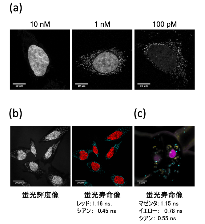
Figure 4: Organellar DNA staining by Kakshine and differentiation from nuclear DNA by fluorescence lifetime imaging
(a) Kakshine selectively stains nuclear DNA at a concentration of 10 nM, mt-DNA at 100 pM, and allows for observation of both at an intermediate concentration of 1nM.
(b) At the concentration that dyes both nuclear and mt-DNA, the two types of DNA can be differentiated using fluorescence lifetime imaging.
(c) Kakshine enables the observation of the three types of plant cell DNA (nu-DNA, mt-DNA, and chl-DNA) with just a single dye, also by fluorescence lifetime imaging. The nu-DNA, mt-DNA, and chl-DNA are shown in magenta, cyan, and yellow, respectively.
Moving on, the group focused on the development of a variant of Kakshine for use in super-resolution STED imaging, a kind of microscopy with resolution beyond the diffraction limit of light. When they combined Kakshine with super-resolution STED imaging, they found that they were able to discern fine structures of cellular DNA and mitochondrial nucleoids (mt-nucleoids), which is not possible with regular confocal microscopy (Figure 5). They were able to estimate from the images that the size of the mitochondrial nucleoids was about 100 nm, which matches previously reported cases. With previously available DNA probes being incompatible with super-resolution STED imaging of mt-nucleoids in living cells, this achievement further highlights the exceptional utility of Kakshine.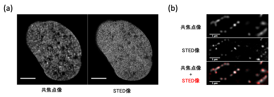
Figure 5: STED imaging
Using super-resolution STED imaging with Kakshine, it was possible to make out the fine structures of nuclear DNA (nu-DNA) and mitochondrial nucleoids (mt-nucleoids), a feat impossible with confocal microscopy.
In summary, Kakshine is an exceptional new series of DNA staining probe which improves upon the capabilities of and solves the shortcomings of current-generation fluorescent dyes such as Hoechst and SYBR-Green 1. It is capable of staining DNA in a variety of animal cells, plant leaf and root cells, without requiring phototoxic UV light for imaging, making it a useful DNA probe in many organisms. It is compatible with a variety of cutting-edge imaging techniques, including two-photon excitation microscopy and super-resolution STED imaging, and when used in conjunction with fluorescence lifetime imaging, it enables the separate observation of nu-DNA, mt-DNA and chl-DNA with just one dye. The medical and life science fields require DNA staining probe for various DNA research methods as well as bioimaging, such as electrophoresis, quantitative PCR and flow cytometry. Kakshine's high DNA selectivity and visible light spectrum capabilities make it ideal for a wide variety of different applications, and it is expected to find use across medicine and life sciences.
Journal Information
The article 'N-aryl pyrido cyanine derivatives are nuclear and organelle DNA markers for two-photon and super-resolution imaging' by Kakishi Uno, Nagisa Sugimoto and Yoshikatsu Sato is published in Nature Communications
2021-05-19