Example of confocal microscopy by Leica TCS-SP8
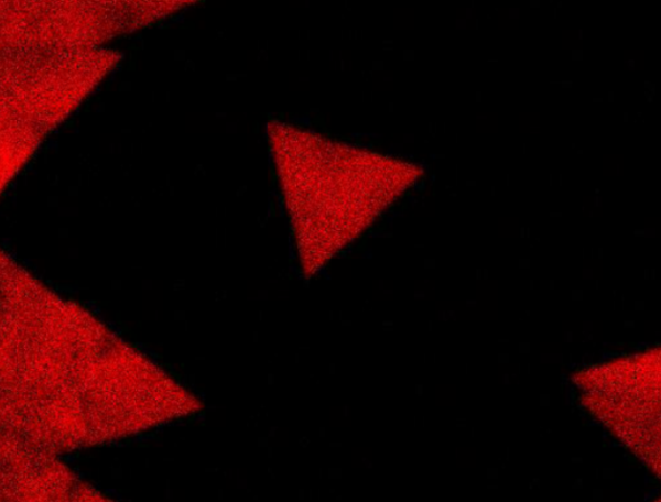
2015/03/23Photoluminescence image of tungsten disulfide(WS2) atomic layer. It can be observed because WS2 atomic layer has direct band gap. Photo by Mitsuhiro Okada (Prof. Shinohara lab.) at Nagoya University.
-

Example of confocal microscopy by Leica TCS-SP8
2015/03/23Photoluminescence image of tungsten disulfide(WS2) atomic layer. It can be observed because WS2 atomic layer has direct band gap. Photo by Mitsuhiro Okada (Prof. Shinohara lab.) at Nagoya University.

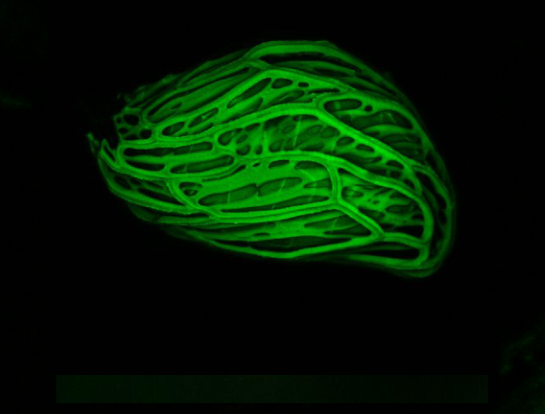
Example of confocal microscopy by LSM780
2015/03/09Seed coat autofluorescence of Striga hermonthica, known as a wichweed. Striga is a parasitic plant and causes serious damage to agriculture in parts of Africa. Photo by Masahiko Yoshimura (Prof. Itami Group), Yuichiro Tsuchiya (Prof. Kinoshita Group) and Yoshikatsu Sato (Live Imaging Center) at ITbM, Nagoya University.

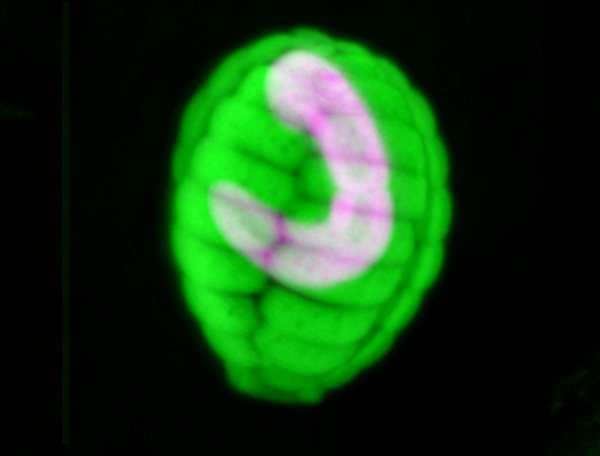
Example of confocal microscopy by Leica TCS-SP8
2015/03/09Shell stained with LysoTracker Yellow HCK-123 (green) and autofluorescence of chromatophore (magenda) in a testate amoeba Paulinella chromatophora (euglyphid). Photo by Mami Nomura at Tsukuba University and Yoshikatsu Sato at ITbM Live Imaging Center.

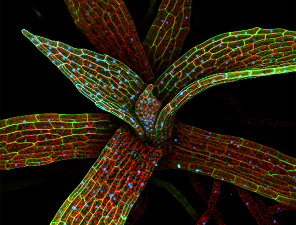
Example of spectral imaging and linear unmixing by LSM780
2015/02/19The cell walls of the moss gametophore leaf cells in the Histon-H2B-mRFP line were stained with propidium iodite (PI). The mRFP, PI and chlorophyll were excited with a 560 nm laser line and collecting 561-677 nm spectrum. Automatic component extraction (ACE) was used for linear unmixing. Photo by Nagisa Sugimoto and Yoshikatsu Sato at ITbM Live Imaging Center, Nagoya University.

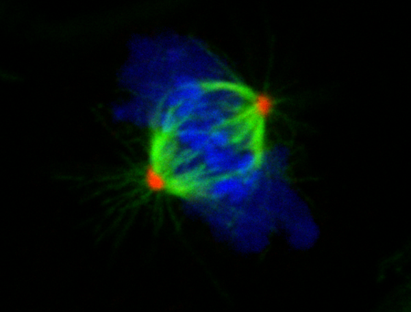
Example of confocal microscopy by Leica TCS-SP8
2015/02/19Dividing NIH 3T3 cells were fixed and stained with anti-gamma-tubulin(red:Dylight649), alpha-tubulin(green: TRITC), and DNA(blue: Hoechst 33342). Photo by Sadanori Watanabe at Nagoya University.